
Table of Contents
Synonyms
“Haemorrhagic septicaemia – Huppe 1886, avian pasteurellosis
Cause
Pasteurella multocida (G-ve, non-motile, non-spore forming, rod 0.2 -0.4 µm by 0.6 – 2.5 µm, aerobic or anaerobic). Classified according to capsular antigens (A, B, D, E and F) by a passive haemaggluitnation test and the tube agglutination or preciptin tests to detect the somatic antigens (serovars 1 to 16). A multiplex capsular PCR assay can now recognise all these serotypes. All except capsular antigen E and somatic antigens 8 and 13 have been found in birds. Capsular antigen A with somatic antigens 3 and 4 are most commonly reconised in the UK and USA associated with virulent Fowl Cholera.
Host
In commercial poultry the turkey is the most sensitive to infection. In chickens, the adult is more sensitive than the juvenile. ducks, geese, pheasants and partridge are also highly susceptible. Infection has also been recorded in birds in zoologiczl collections and wild birds. It is likely that most species of birds can be infected. Pigs, cats and rodents can be carriers that will cause disease in birds.
Transmission
P.multocida primarily infects the respiratory tract but can then spread down to the lungs before becoming bacteraemic and septicaemic. Lateral spread is mainly through discharges from the mouth, nose and eyes which contaminate feed and water. It is rarely found in the faeces. Infection can occur through cuts in the skin and be acquired from eating carcases. Chronic carrier birds and rodents are important sources of infection. On contaminated pasture, disease most commonly appears after wet weather.
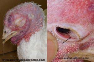
Clinical signs
Presents in two forms – acute and chronic. The presence of a capsule is associated with virulence whilst endotoxins will exacerbate signs. Acute infection: – If not found dead, respiratory distress, fevered, ruffled feathers, anorexia and lethargy may be seen. Acute – reddening and cyanosis of skin of head, mucus from the beak and closed eyes. Chronic infection:- swollen wattles and swelling between the wattles. Occasionally torticollis due to a meningitis. Chronic – Tracheal rales, infectious arthritis of legs and wings. No specific egg abnormalities in laying flocks. Torticollis may be seen where infection of the middle ear, skull and meninges occurs.
Pathology
Acute – petechial haemorrhages on the heart, abdominal fat, a consolidated pneumonia, congestion and miliary pinpoint foci in the liver, sticky mucus in intestinal tract and a generalised septicaemia (bloody discolouration of the abdominal contents). Chronic – swelling of the face and wattles, peritonitis, salpingitis, infectious arthritis and sternal bursitis.
Diagnosis
The history and clinical signs will indicate the possibility of Fowl Cholera. In acute cases bacteria can be isolated from viscera on blood agar, aerobic incubation for 24 – 48 hours at 37oC. Impression smears stained with Methylene Blue will reveal typical safety pin shaped bacteria. PCR can also be used to detect P.multocida in pure and mixed cultures or from clinical samples.
Differential diagnoses
Acute: Newcastle disease, Avian Influenza, ILT, Erysipelas, Colibacillosis, Salmonella gallinarum, Avibacterium gallinarum. Chronic: Staphylococcal infections, ORT.
Treatment
Antibiotic therapy can help e.g. Amoxycillin, tetracyclines and fluoroquinolones. this can be combined with vaccination in the face of severe outbreaks. There is a wide variation in the response to both treatment and vaccination. Relapse after treatment is common particularly in free range egg laying flocks on ground previously used by infected pig herds.
Prevention
P.multocida can adapt to evase the hosts immune system. Hence vaccination does not offer total protection. Consequently, improved control may be obtained by using both licenced and autogenous vaccines. In the UK, Poulvac Pabac IV is available. Particular attention should be paid to rodent control, mixing with other farm animals and remember pets can introduce infection along with the purchase of carrier birds.
Fowl cholera is the 15th commonest diagnosis made by my local AHVLA labs. Details of species affected, clinical signs and post mortem lesions observed can be found by clicking here.